Lab
1: Principles and Use of Microscope
1.1
Setting up and using the microscope
Introduction:
A microscope is an instrument used to see objects
that are invisible to the naked eye. The science of investigating small objects
using such an instrument is called microscopy. Microscopic means invisible to
the eye unless aided by a microscope. There are many types of microscopes. The
most common use is the optical microscope (light microscope), which uses
visible light and a system of lenses to magnify images of small samples.
The diagram below shows the parts of the microscope:

STRUCTURAL COMPONENTS
OF A MICROSCOPE
The three basic,
structural components of a compound microscope are the head, base and arm.
• Head/Body: Houses the optical parts
in the upper part of the microscope
• Base: Supports the microscope and
houses the illuminator
• Arm: Connects to the base and
supports the microscope head. It is also used to carry the microscope.
OPTICAL COMPONENTS OF A
MICROSCOPE
There are two optical
systems in a compound microscope: Eyepiece Lenses and Objective Lenses:
· Eyepiece
or Ocular: The lens at the top that you look through.
Typically, standard eyepieces have a magnifying power of 10x.
·
Eyepiece
Tube:
Holds the eyepieces in place above the objective lens. Binocular microscope
heads typically incorporate a diopter adjustment ring that allows for the
possible inconsistencies of our eyesight in one or both eyes. Binocular
microscopes also swivel (Interpupillary Adjustment) to allow for different
distances between the eyes of different individuals.
·
Objective
Lenses: The primary optical lenses on a microscope. They
range from 4x-100x and typically, include, three, four or five on lens on most
microscopes.
·
Nosepiece:
Holds two or more objective lenses and can be rotated to easily change power.
·
Coarse
focus knob: It is used to focus on the specimen. It may move
either the stage or the upper part of the microscope in a relative up and down
motion.
·
Fine
Focus knobs: It is the smaller round knob on the
side of the microscope used to fine-tune the focus of the specimen after using
the coarse adjustment knob.
·
Stage
Clips: Used when there is no mechanical stage. The viewer
is required to move the slide manually to view different sections of the
specimen.
·
Aperture:
The hole in the stage through which the base (transmitted) light reaches the
stage.
·
Illuminator:
The light source for a microscope, typically located in the base of the
microscope.
·
Iris
Diaphragm: Controls the amount of light reaching the
specimen. It is located above the condenser and below the stage.
·
Condenser
Focus Knob: Moves the condenser up or down to control the
lighting focus on the specimen.
Magnification
and resolution:
Magnification is the ability to make small objects seem
larger, such as making a microscopic organism visible. There are four
magnifications in the microscope used:
4x objective X 10x
eyepiece = 40x magnification
10x objective X 10x
eyepiece = 100x magnification
40x objective X 10x
eyepiece = 400x magnification
100x objective X 10x
eyepiece = 1000x magnification
Resolution is the ability to distinguish two objects from
each other. Not to be confused with magnification, microscope resolution is the
shortest distance between two separate points in a microscope’s field of view
that can still be distinguished as distinct entities.
Objective:
-To learn the proper
way of handling and care of microscope.
-To understand the
basic concept of magnification and resolution of a microscope.
Materials
and reagents:
-Microscope slide and
cover-slip
Procedure:
(Refer to the lab
manual)
Results


Discussion
1.2 Examination of cells
Introduction
Living microorganisms are very diverse in their type, size and shape but most importantly many of them are also colorless when viewed using the microscope. It is essential as a biology student that we can search and found easily the microorganisms that is used so that we can examine and learn from it.
As such, by using the wet mount methods which does not require any special equipment, not harmful when done properly and significantly quick and easy we can studies more on microorganism easier. The wet mount methods are a technique that enable us to experience the proper use of microscope and aseptically prepare a slide thus, we are learning in a safe environment.
Objective
-To provide an experience in the use of microscope
-To illustrate the diversity of cells and microorganisms
Materials and reagents
-Culture
-Immersion Oil
-Lens Tissue
-A microscope slide containing stained microorganisms
-Inoculating loop
-Bunsen burner
-Slide and coverslip
Result

Discussion
References
Results

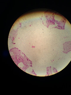
Typical Bacillus is observed under the microscope

40x magnification

100x magnification

400x magnification

1000x maginification (oil immersion)
We observed the specimen from lowest magnification that
is 40x to the highest magnification 1000x (oil immersion). The specimen that we
observed is Typical Bacillus. Bacillus is a genus of Gram-positive
which appeared pink in color when observed under the microscope. Bacillus is one of the best understood
prokaryotes, in terms of molecular and cellular biology. The cell wall of Bacillus is a structure on the outside
of the cell that forms the second barrier between the bacterium and the
environment and at the same time maintains the rod shape and withstands the
pressure generated by the cell’s turgor. The cells are straight, round-ended or
square-ended rods. The size of Typical
Bacillus is tiny and the surface as it was observed under the magnification
of 1000x with oil immersion, it looked smooth and the texture is moist. Most
species motile by peritrichous flagella. Many Bacillus species have little or no pathogenicity and are rarely
associated with disease in humans or lower animals except Bacillus anthracis, Bacillus cereus and Bacillus subtilis. Some species are insect pathogens.
Conclusion
In conclusion, we have learnt the right way of handling
the microscope with care and also how to observe a specimen under the
microscope from the lowest magnification to the highest magnification using a
correct way. We find that the higher the magnification, the clearer the image
that we can see under the microscope.
1.2 Examination of cells
Introduction
Living microorganisms are very diverse in their type, size and shape but most importantly many of them are also colorless when viewed using the microscope. It is essential as a biology student that we can search and found easily the microorganisms that is used so that we can examine and learn from it.
As such, by using the wet mount methods which does not require any special equipment, not harmful when done properly and significantly quick and easy we can studies more on microorganism easier. The wet mount methods are a technique that enable us to experience the proper use of microscope and aseptically prepare a slide thus, we are learning in a safe environment.
Objective
-To provide an experience in the use of microscope
-To illustrate the diversity of cells and microorganisms
Materials and reagents
-Culture
-Immersion Oil
-Lens Tissue
-A microscope slide containing stained microorganisms
-Inoculating loop
-Bunsen burner
-Slide and coverslip
Result

Lactobacillus (1000x magnification with oil immersion)
Discussion
The wet mount methods enable us to observe the
Lactobacillus more clearly of the sizes and the shape. Oil immersion fills the space
between the objective lens and specimen and matches the refractive index of the
glass cover slip and glass objective lens. At a given focal length, greater
numerical aperture can be achieved.
Lactobacillus is a genus of Gram-positive facultative
anaerobic or rod-shaped bacteria. They are a major part of the lactic acid
bacteria group. In humans they are part of the vaginal microbiota.
Lactobacillus is a type of bacteria with multiple different species in the
genus. Most Lactobacillus species in humans are considered harmless.
Lactobacilli live in the urinary digestive and genital tracks of humans.Some
Lactobacillus species are used as starter culture in industry for controlled
fermentation in the production of yogurt, cheese, beer, wine etc.
In this experiment, aseptic techniques is important to
ensure that the bacteria is not contaminated. It is also to protect the user
from infection and to prevent the spread of microorganisms. Through this
experiment, Lactobacillus appeared to be translucent because it was not
stained.
Conclusion
The bacteria can be seen more clearly with the highest
magnification under oil immersion. Added with wet mount method, the bacteria
can be seen clearly in its natural state. Proper aseptic technique is important
because some of the pathogenic agents can cause infection or serious illness to
the user if they are not handled in a proper way.
References
https://en.wikipedia.org/wiki/Microscope
http://www.microscope.com/education-center/microscopes-101/compound-microscope-parts/
http://sciencelearn.org.nz/Contexts/Exploring-with-Microscopes/Science-Ideas-and-Concepts/Magnification-and-resolution
www.en.m.wikipedia.org/wiki/bacillus.com
No comments:
Post a Comment